You provide care for quite a few people who have knee pain. Sometimes you see these patients before surgery – other times after surgery. When someone has persistent knee pain, what is something you typically note during your examination?
For me, most of my patients who are receiving services for long standing pain in their knee tend to have gait abnormalities. Sometimes their gait is slow…. sometimes they have an obvious limp. My role is not only to help with their pain experience, but to also improve their level of function.
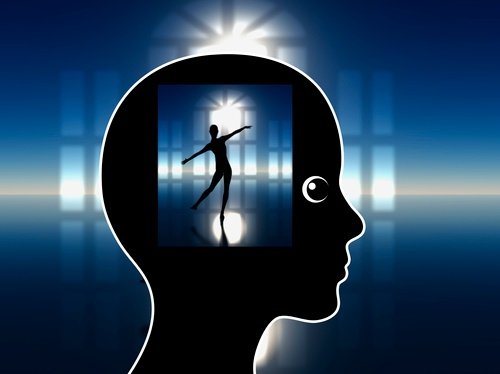
What this study is so great at outlining is the neuroplasticity of the brain. Those who have long standing gait abnormalities demonstrate changes in their brain to such a degree that the brain no longer has the capability to imagine a normal movement pattern.
This means that motor imagery will not be the best cognitive tool in your box. Your brain will do better with visual imagery – where the patient watches the desired activity. This makes sense because when I ask patients to think about a particular movement and how it feels, it’s almost as though they have a disconnect. If I use a video or my hands… or even what my patients call my “Stride Rite” treadmill, they immediately understand and are able to attempt to change their preferred, learned over time, movement patterns.
This research may also help explain why after a person has surgery and no longer has pain that the abnormal gait characteristics persist. The neuroplasticity of the brain created new movement maps and maintained that map, even after surgery and pain relief.
You’ll find the abstract to the recent study below.
A functional limitation to the lower limbs affects the neural bases of motor imagery of gait.
Abstract
Studies on athletes or neurological patients with motor disorders have shown a close link between motor experience and motor imagery skills. Here we evaluated whether a functional limitation due to a musculoskeletal disorder has an impact on the ability to mentally rehearse the motor patterns of walking, an overlearned and highly automatic behaviour.
We assessed the behavioural performance (measured through mental chronometry tasks) and the neural signatures of motor imagery of gait in patients with chronic knee arthrosis and in age-matched, healthy controls. During fMRI, participants observed (i) stationary or (ii) moving videos of a path in a park shown in the first-person perspective: they were asked to imagine themselves (i) standing on or (ii) walking along the path, as if the camera were “their own eyes” (gait imagery (GI) task). In half of the trials, participants performed a dynamic gait imagery (DGI) task by combining foot movements with GI.
Behavioural tests revealed a lower degree of isochrony between imagined and performed walking in the patients, indicating impairment in the ability to mentally rehearse gait motor patterns. Moreover, fMRI showed widespread hypoactivation during GI in motor planning (premotor and parietal) brain regions, the brainstem, and the cerebellum. Crucially, the performance of DGI had a modulatory effect on the patients and enhanced activation of the posterior parietal, brainstem, and cerebellar regions that the healthy controls recruited during the GI task.
These findings show that functional limitations of peripheral origin may impact on gait motor representations, providing a rationale for cognitive rehabilitation protocols in patients with gait disorders of orthopaedic nature. The DGI task may be a suitable tool in this respect.
Neuroimage Clin. 2018 Jul 5;20:177-187. doi: 10.1016/j.nicl.2018.07.003. eCollection 2018.
{{cta(‘c6ecd159-44e1-4eb3-9940-fbb5cb2761d5’)}}
Leave a Reply